
Donkey Secondary Antibodies


Biotium Primary Antibody Human IgG Fc N2508.HFC CF647 1/EA
Anti-Human IgG (Fc) rVHH (N2508 HFC) is a recombinant Alpaca VHH antibody that recognizes the Fc domain of human IgG and is validated for IF and flow Far-red fluorescent STORM-compatible CF647 dye has Ex/Em at 650/665 nm Unit size 500 uL at 0 1 mg/mL


Biotium Primary Antibody Rabbit IgG Fc N2507.RFC CF647 1/EA
Alpaca Anti-Rabbit IgG (Fc) rVHH (N2507 RFC) is a recombinant Alpaca VHH antibody that recognizes the Fc fragment of rabbit IgG and is validated for immunofluorescence Far-red fluorescent STORM-compatible CF647 dye has Ex/Em at 650/665 nm Unit size 500 uL at 0 1 mg/mL

Biotium VLDL-Receptor (Very Low Density Lipoprotein Receptor) (VLDLR/1337), CF647 conjugate, 0.1mg/mL
VLDLR (very low density lipoprotein receptor) is a member of the LDL receptor gene family, which includes LDL receptor, LRP, megalin, VLDLR and ApoER2. The LDL receptor family is characterized by a cluster of cysteine-rich class A repeats, epidermal growth factor (EGF)-like repeats, YWTD repeats and an O-linked sugar domain. VLDLR associates with RAP (receptor associated protein) during receptor folding, and RAP facilitates the secretion of the extracellular region of VLDLR. VLDLR is thought to mediate the interaction of extracellular Reelin and cytosolic mDab1 (mammalian disabled protein), which activates a tyrosine kinase. This pathway regulates the migration of neurons along the radial glial fiber network during brain development.Primary antibodies are available purified, or with a selection of fluorescent CF Dyes and other labels. CF Dyes offer exceptional brightness and photostability. Note: Conjugates of blue fluorescent dyes like CF405S and CF405M are not recommen
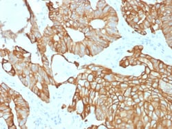
Biotium Cytokeratin 19 (Pancreatic Stem Cell Marker) (rKRT19/800), CF594 conjugate, 0.1mg/mL
This antibody recognizes a protein of 40 kDa, identified as cytokeratin-19 (CK19), which is expressed in sweat gland, mammary gland ductal and secretory cells, bile ducts, gastrointestinal tract, bladder urothelium, oral epithelia, esophagus, and ectocervical epithelium. Anti-CK19 reacts with a wide variety of epithelial malignancies including adenocarcinomas of the colon, stomach, pancreas, biliary tract, liver, and breast. Perhaps the most useful application is the identification of thyroid carcinoma of the papillary type, although 50%-60% of follicular carcinomas are also labeled. Anti-CK19 is a useful marker for detection of tumor cells in lymph nodes, peripheral blood, bone marrow and breast cancer.Primary antibodies are available purified, or with a selection of fluorescent CF Dyes and other labels. CF Dyes offer exceptional brightness and photostability. Note: Conjugates of blue fluorescent dyes like CF405S and CF405M are not recommended for detecting low abundanc
Biotium Glypican-3 (GPC3) (Hepatocellular Carcinoma Marker) (rGPC3/863), CF647 conjugate, 0.1mg/mL
Glypican-3 (GPC3) is a glycosylphospatidyl inositol-anchored membrane protein, which may also be found in secreted form. Anti-GPC3 has been identified as a useful tumor marker for the diagnosis of hepatocellular carcinoma (HCC), hepatoblastoma, melanoma, testicular germ cell tumors, and Wilm s tumor. In patients with HCC, GPC3 is overexpressed in neoplastic liver tissue and elevated in serum, but is undetectable in normal liver, benign liver, and the serum of healthy donors. GPC3 expression is also found to be higher in HCC liver tissue than in cirrhotic liver or liver with focal lesions such as dysplastic nodules and areas of hepatic adenoma (HA) with malignant transformation. In the context of testicular germ cell tumors, GPC3 expression is up regulated in certain histologic subtypes, specifically yolk sac tumors and choriocarcinoma. A high level of GPC3 expression is also found in some types of embryonal tumors, such as Wilm s tumor and hepatoblastoma, with a low or undete
Biotium CD257 / BAFF / TNFSF13B (C257/1638), CF647 conjugate, 0.1mg/mL
BAFF/CD257 is a type II transmembrane protein, and a member of the tumor necrosis factor ligand superfamily. It is proteolytically cleaved to form a soluble protein. BAFF, like other TNF ligand family members, forms homo-trimers. The predicted molecular weight of a BAFF monomer is approximately 31 kDa. It is expressed on peripheral blood B and T lymphocytes, monocytes, macrophages, and dendritic cells and is upregulated by IFN-gamma and down-regulated by PMA/ionomycin treatment. BAFF stimulates B and T cell immunity and regulates humoral immunity by binding TACI and BCMA receptors; BAFF prevents apoptosis and promotes B cell survival by binding the BAFF-specific receptor (BAFFR/BR3).Primary antibodies are available purified, or with a selection of fluorescent CF Dyes and other labels. CF Dyes offer exceptional brightness and photostability. Note: Conjugates of blue fluorescent dyes like CF405S and CF405M are not recommended for detecting low abundance targets, because
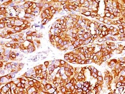
Biotium MART-1 / Melan-A / MLANA (Melanoma Marker) (MLANA/1409R), CF647 conjugate, 0.1mg/mL
This antibody recognizes a protein doublet of 20-22 kDa, identified as MART-1 (Melanoma Antigen Recognized by T cells 1) or Melan-A. MART-1 is a newly identified melanocyte differentiation antigen recognized by autologous cytotoxic T lymphocytes. Seven other melanoma associated antigens recognized by autologous cytotoxic T cells include MAGE-1, MAGE-3, tyrosinase, gp100, gp75, BAGE-1, and GAGE-1. Subcellular fractionation shows that MART-1 is present in melanosomes and endoplasmic reticulum. This MAb labels melanomas and other tumors showing melanocytic differentiation. It is also a useful positive-marker for angiomyolipomas. It does not stain tumor cells of epithelial, lymphoid, glial, or mesenchymal origin.Primary antibodies are available purified, or with a selection of fluorescent CF Dyes and other labels. CF Dyes offer exceptional brightness and photostability. Note: Conjugates of blue fluorescent dyes like CF405S and CF405M are not recommended for detecting low a
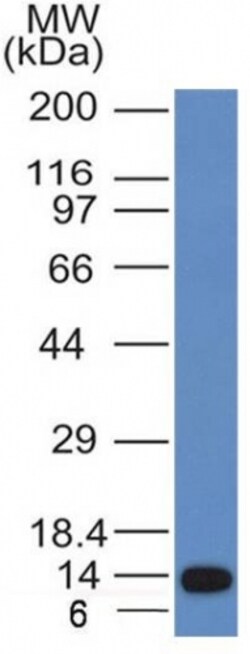
Biotium ALK (Anaplastic Lymphoma Kinase) / CD246(ALK/1031), CF647 conjugate, 0.1mg/mL
The wild-type anaplastic lymphoma kinase (ALK) protein is a 200 kDa transmembrane receptor tyrosine kinase. Its expression is restricted to a few scattered cells in the nervous system (some glial cells and neurons, and a few endothelial cells and pericytes). ALK has been found to be rearranged, mutated, or amplified in a series of tumors. The hybrid gene, NPM-ALK, created by the t(2;5)(p23;q35) chromosomal translocation, encodes part of the nucleolar phosphoprotein nucleophosmin (NPM), joined to the entire cytoplasmic portion of ALK. As a consequence, the ALK gene comes under the control of the NPM promoter, which induces a permanent and ubiquitous transcription of the NPM-ALK hybrid gene, resulting in the production of a 80 kDa NPM-ALK chimeric protein. This translocation is found in anaplastic large cell lymphomas (ALCL). Reportedly, expression of ALK indicates a better prognosis. Approximately 5%-10% of non-small cell lung carcinomas also express ALK protein, producing a c
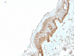
Biotium S100A2 / S100 Calcium Binding Protein A2(CPTC-S100A2-2), CF647 conjugate, 0.1mg/mL
S100A2 belongs to the S100 family of proteins containing 2 EF-hand calcium-binding motifs. S100A2 may function as a modulator against excess calcium accumulation in normal human mammary epithelial cells and also have a role in suppressing tumor cell growth. This protein may have a tumor suppressor function. Chromosomal rearrangements and altered expression of this gene have been implicated in breast cancer. Cytoplasmic overexpression may also be of prognostic significance when observed in oral cancer patients, and the S100A2 has also been identified as significantly down-regulated in gastric cancer. Primary antibodies are available purified, or with a selection of fluorescent CF Dyes and other labels. CF Dyes offer exceptional brightness and photostability. Note: Conjugates of blue fluorescent dyes like CF405S and CF405M are not recommended for detecting low abundance targets, because blue dyes have lower fluorescence and can give higher non-specific background than othe
Biotium Human IgG (Immunoglobulin Gamma Heavy Chain) (B-Cell Marker) (rIG266), CF647 conjugate, 0.1mg/mL
Recognizes a protein of 75 kDa, identified as gamma heavy chain of human immunoglobulins. It does not cross-react with alpha (IgA), mu (IgM), epsilon (IgE), or delta (IgD), heavy chains, T-cells, monocytes, granulocytes, or erythrocytes. This MAb is useful in the identification of leukemias, plasmacytomas, and certain non-Hodgkin's lymphomas. The most common feature of these malignancies is the restricted expression of a single heavy chain class. Demonstration of clonality in lymphoid infiltrates indicates that the infiltrate is clonal and therefore malignant. Primary antibodies are available purified, or with a selection of fluorescent CF Dyes and other labels. CF Dyes offer exceptional brightness and photostability. Note: Conjugates of blue fluorescent dyes like CF405S and CF405M are not recommended for detecting low abundance targets, because blue dyes have lower fluorescence and can give higher non-specific background than other dye colors.
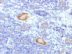
Biotium von Willebrand Factor / Factor VIII Related-Ag (Endothelial Marker) (VWF/1859R), CF647 conjugate, 0.1mg/mL
von Willebrand Factor (vWF) is a multimeric glycoprotein that is found in endothelial cells, plasma and platelets. It acts as a carrier protein for Factor VIII and promotes platelet adhesion and aggregation. vWF undergoes a variety of posttranslational modifications that influence the affinity and availability for Factor VIII, including cleavage of the propeptide and formation of N-terminal disulfide bonds. This antibody helps to establish the endothelial nature of some lesions of disputed histogenesis, e.g. Kaposi's sarcoma and cardiac myxoma. It is widely used for differentiating vascular lesions from those of other tissue differentiation within a panel of other vascular markers although not all tumors of endothelial differentiation contain this antigen.Primary antibodies are available purified, or with a selection of fluorescent CF Dyes and other labels. CF Dyes offer exceptional brightness and photostability. Note: Conjugates of blue fluorescent dyes like CF405S and C
Biotium E-Cadherin / CD324 (Intercellular Junction Marker) (CDH1/1525), CF647 conjugate, 0.1mg/mL
This antibody recognizes a protein of 120-80 kDa, identified as E-cadherin. Cadherins comprise a family of Ca2 -dependent adhesion molecules that function to mediate cell-cell binding critical to the maintenance of tissue structure and morphogenesis. The classical cadherins, E-, N- and P-cadherin, consist of large extracellular domains characterized by a series of five homologous NH2 terminal repeats. The relatively short intracellular domains interact with a variety of cytoplasmic proteins, such as β-catenin, to regulate cadherin function. E-cadherin plays an important role in epithelial cell adhesion. A decreased expression of E-cadherin is associated with metastatic potential and poor prognosis in breast cancer, prostate and esophageal cancer. In combination with p120 Catenin, it is useful for the differentiation between ductal (E-cadherin ) and lobular (E-cadherin -) breast carcinomas. It may also help in diagnosis of mesothelioma.Primary antibodies are available
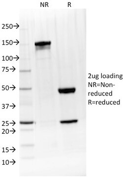
Biotium IL-4 / Interleukin 4(IL4/1597), CF647 conjugate, 0.1mg/mL
Interleukin-4 (IL-4), also designated B cell stimulatory factor-1, is a glycosylated cytokine secreted by activated T lymphocytes, basophils and mast cells. The secreted IL-4 protein promotes the growth and differentiation of cells that participate in immune defense by favoring such events as the expansion of the Th2 lineage relative to Th1 cells. These T helper cell subsets are defined by their pattern of cytokine secretion: Th1 cells secrete IL-2, TNF, while Th2 cells secrete IL-4, IL-5 and IL-10. Another key immunological function of IL-4 is to induce immunoglobulin class switching. IL-4 has been shown to induce the production of IgE and enhance IgG4 secretion by B cells, but suppress the production of IgM, IgA, IgG1, IgG2 and IgG3. It has been determined that Stat6 is rapidly tyrosine phosphorylated following stimulation of IL-3 or IL-4, but is not detectably phosphorylated following stimulation with IL-2, IL-12 or erythropoietin.Primary antibodies are available purif
Biotium Cytokeratin, Acidic (Type I or LMW) (Epithelial Marker) (KRTL/1377), CF488A conjugate, 0.1mg/mL
This MAb recognizes the 56.5 kDa (CK10); 50 kDa (CK14); 50 kDa (CK15); 48 kDa (CK16); 40 kDa (CK19) keratins of the acidic (Type I or LMW) subfamily. Twenty human keratins are resolved with two-dimensional gel electrophoresis into acidic (pI 6.0) subfamilies. The acidic keratins have molecular weights (MW) of 56.5, 55, 51, 50, 50 , 48, 46, 45, and 40 kDa. Many studies have shown the usefulness of keratins as markers in cancer research and tumor diagnosis.Primary antibodies are available purified, or with a selection of fluorescent CF Dyes and other labels. CF Dyes offer exceptional brightness and photostability. Note: Conjugates of blue fluorescent dyes like CF405S and CF405M are not recommended for detecting low abundance targets, because blue dyes have lower fluorescence and can give higher non-specific background than other dye colors.
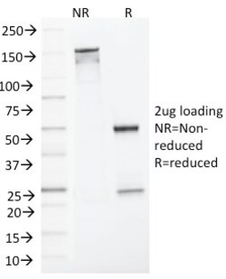
Biotium MLH1 (MutL Homolog 1)(MLH1/1324), CF647 conjugate, 0.1mg/mL
This MAb recognizes a protein of 83 kDa, identified as MLH1. Defects in MLH1 are the cause of hereditary non-polyposis colorectal cancer type 2 (HNPCC2). Heterodimerizes with PMS2 to form MutL alpha, a component of the post-replicative DNA mismatch repair system (MMR). DNA repair is initiated by MutS alpha (MSH2-MSH6) or MutS beta (MSH2-MSH6) binding to a dsDNA mismatch, then MutL alpha is recruited to the heteroduplex. Assembly of the MutL-MutS-heteroduplex ternary complex in presence of RFC and PCNA is sufficient to activate endonuclease activity of PMS2. It introduces single-strand breaks near the mismatch and thus generates new entry points for the exonuclease EXO1 to degrade the strand containing the mismatch. DNA methylation would prevent cleavage and therefore assure that only the newly mutated DNA strand is going to be corrected. MutL alpha (MLH1-PMS2) interacts physically with the clamp loader subunits of DNA polymerase III, suggesting that it may play a role to recr